New, non-destructive imaging techniques are being used to gain a better understanding of the impact of the fungi involved in grapevine trunk decay phenomena.
The Vitimage project aims to assess the potential of imaging tools to study and detect grapevine trunk diseases. “More specifically, it involves using non-destructive imaging tools, known in particular for their use in medical applications, to monitor the development and impact of fungi on grapevine trunks, and try to detect them without waiting for the possible expression of leaf symptoms,” says Cédric Moisy of IFV, who heads up the Vitimage project. The aim is to complement existing knowledge about grapevine trunk diseases and to evaluate the impact of pathogenic fungi on woody tissues more precisely. However, when talking about this exploratory project, which began in early 2018 and will run for 3 years, Cédric Moisy explains that “the first step is to evaluate and assess the relevance of imaging tools, in order to determine whether they can be used to detect and monitor grapevine trunk diseases”.
Characterizing the symptoms and impact of each fungus
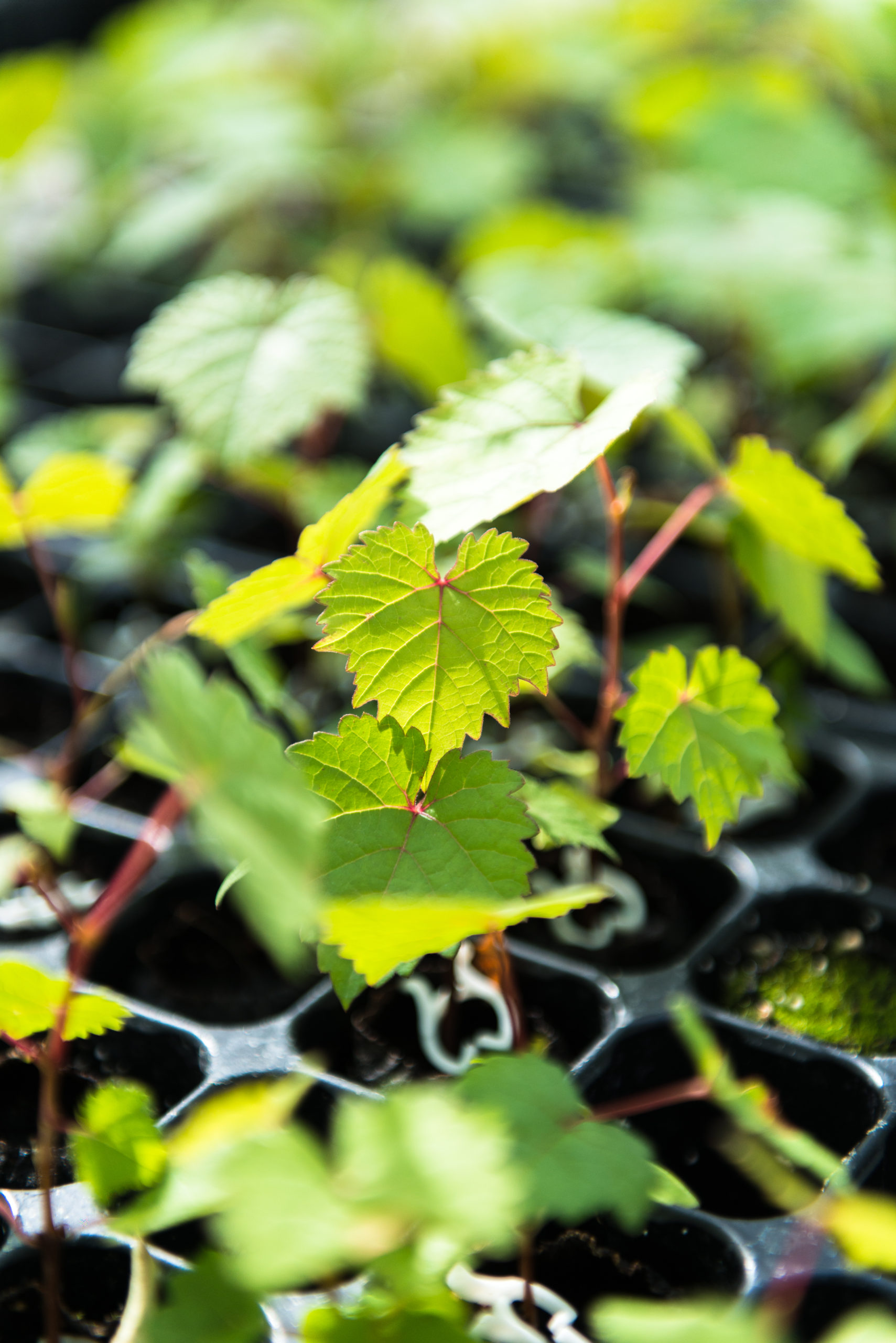
“We are implementing two complementary approaches. On the one hand, we work on cuttings inoculated under controlled conditions with known pathogens, which we study in micro-imaging over an area of 2 to 4 cm around the inoculation point.
The objectives of this first approach are to test the tools and to describe and compare the spread of the different fungi tested and to identify new disease markers.
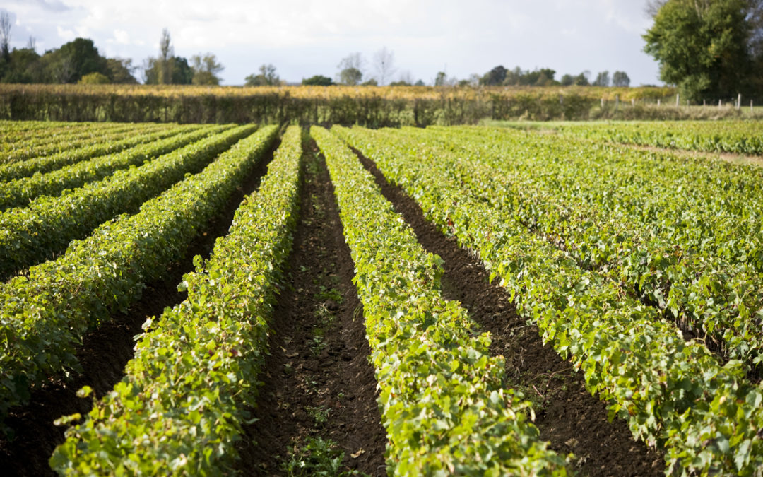
In addition, studies are being conducted with the CIVC (Comité Interprofessionnel du Vin de Champagne) on vines with different disease expression profiles, taken from a study plot in the Champagne vineyards. “At the beginning of the year, Julie Perry (CIVC) took samples from three types of individual: ‘asymptomatic’ (i.e. had never expressed symptoms), ‘symptomatic’ (expressing symptoms in the year of collection), and finally ‘resilient’ (expressing symptoms in previous years, but not in the year of collection),” says Cédric Moisy. The vines are then taken to the Radiology and Physiotherapy Centre (CRP) at Clinique du Parc at Castelnau-Le-Lez in Greater Montpellier, to be scanned by Dr Samuel Mérigeaud using an MRI machine or an X-ray scanner normally used for humans. Sessions are generally held at night so as not to disrupt the operation of the service. Further technical tests are envisaged in the project. “We haven’t tested the spectrometry yet. As for ultrasound or ultrasound imaging, it is not really suitable for this type of examination,” adds Cédric Moisy. “We are in the calibration phase for these imaging tools.”
- [Webinar] Solutions for alternative weed control #1 Electric weeding – Case studies in vineyard, grassland - 27 April 2023
- Mechanical weeding and required technical skills - 26 March 2023
- 10 questions and answers about plant cover in the vineyard - 25 March 2023
- What are the new avenues of research in system experimentation? - 23 March 2023
- What is clonal selection? - 23 March 2023
- Phthalates : Potential sources and control measures - 23 March 2023
- Rosé wines: impact of storage conditions in tank on the polyphenol composition and color - 29 March 2021
- Reducing alcohol content in wines by combining canopy management practices and biological techniques - 29 March 2021
- Enzymes in oenology : production, regulation, applications - 29 March 2021
- Herbicide-free strategies - 26 March 2021
Réagir à l'article
Pour pouvoir laisser un commentaire, vous devez être inscrit sur notre site.
M'inscrireL'inscription est gratuite.
Déjà inscrit
Lost your password?